Languages
Neuroscience Research
Chemical synapses in the human brain are small.
Due to properties like size and molecular density, synapses and other structures of interest to neuroscience researchers have been difficult to study by conventional methods of microscopic investigation. Yet extracting information about the spatial organization, stoichiometry, interactions, and dynamics of these structures on a molecular level is essential; advancing our understanding of the human brain depends on developing more and better methods to do so.
理想的神经科学显微镜方法不仅应比常规手段实现更高的分辨率,而且还应区分特定的结构,并为多种样本类型提供完整而可靠的信息。
In providing exactly these capabilities, super-resolution microscopy with single-molecule localization (SMLM) has overcome many of the limitations of conventional neuroscience microscopy. This has already enabled dramatic improvement of our understanding of neuronal and brain function and will continue to create new opportunities for investigation and discovery in the field going forward.
Are you interested in Bruker's best-in-class solutions for super-resolution SMLM?
SMLM vs. Other Neuroscience Microscopy Methods
Neuroscience researchers face a unique challenge when selecting an imaging methodology: historically, no single method has been able to provide complete information about neuronal structures and processes at the molecular level, so selecting one has required compromising between capabilities or conducting complementary investigations via multiple methods.
For example, many structures of interest are barely visible or undiscoverable by methods that are capable of specific labeling, like diffraction-limited two-photon microscopy. Yet electron microscopy, which provides superior resolution and is the only method capable of visualizing structures of interest smaller than 50 nm — like the synaptic cleft and synaptic vesicles — lacks fluorescence microscopes' capability for easy protein-labeling of specific structures.1,2This restricts researchers' opportunities to perform complex investigations, including protein identification or neuronal tracing.
How is Super-Resolution SMLM Different?
Contrasting conventional methods of neuroscience microscopy, super-resolution microscopy both enables target-specific fluorescence labeling and achieves resolutions well below the diffraction limit of light microscopy. As a result, it can provide unique and often otherwise inaccessible insights into structures and processes.
在神经科学研究中使用的各种超分辨率成像技术中,包括结构化刷新显微镜(SIM),刺激的发射排放(STED)和扩展显微镜 - SMLM是唯一完全依赖于对分子分析的唯一方法。SMLM also provides the highest resolution.3结果,SMLM的横向分辨率为20 nm和特定的荧光标记,是推进神经科学的精美工具.
Practical Considerations for Super-Resolution SMLM in Neuroscience Applications
What is SMLM Used for in Neuroscience Research?
由于其在标记特异性和解决方案的标签上具有优势,因此SMLM有可能高度适用于神经科学研究人员和SMLM技术的许多感兴趣问题,并且已经在广泛的神经科学应用中使用。
To date, the results of these studies have demonstrated the applicability of the method to both structural and quantitative analysis as well as 3D imaging of both tissue and cultured samples.3In these applications, SMLM has been able to provide insight into the spatial organization, stoichiometry, and temporal dynamics of a broad range of structures and processes and provides significant advantages over other methods for investigating molecular interactions (molecular size, density, distance apart, overlap, etc.).
Does SMLM Have Limitations for Neuroscience Research?
SMLM studies demonstrated on tissue samples appear less frequently in SMLM literature than those demonstrated on cultured cells. This imbalance is especially apparent for applications that call for 3D imaging in tissue, and can be attributed to several sources of frustration for researchers. These include:
- Sample preparation is more complex and time-consuming for tissue samples than for cultured cells
- 组织样品表现出更多(荧光分子)的背景信号更多(更具破坏性)3(并且需要更高级的分析工具来处理)
结果,SMLM技术的可实现的成像深度可能会为使用组织切片和厚样品而构成挑战。
*某些仪器可实现出色的成像深度,尤其是在组织样品中的3D成像
(看 ”Bruker的Vutara VXL在神经科学应用中的优点" for more information).
SMLM适用于神经元结构和过程的研究,包括(其他):
- 神经发展
- Neural circuit assembly
- 突触形成
- 突触传输
- Synaptic proteins
Recommendations for SMLM-Capable Neuroscience Microscopes
What You Need to Know About SMLM for Neuroscience Applications
Depending on the equipment, sample, and fluorophore is used, SMLM methods have the potential to achieve high lateral localization precisions and ~20 nm spatial resolution. Yet the actual resolution and localization precision achieved when imaging both cultured samples and tissue slices depend on a variety of additional factors. These include, among others, labeling density and accuracy, signal size and strength, and microscope drift. Optimizing sample praparation methods for SMLM by, for example, increasing labeling antibody concentration, can resolve many of these issues and clear the way for successful analysis.
为什么选择工具与技术一样重要
SMLM成像系统本身的组件和功能可以限制各种SMLM技术中的任何一种分辨率和本地化精度 -特别是在组织切片中执行3D成像时. Moreover, SMLM-capable microscopes can require time-consuming setup that distracts from critical research processes. They also often pair with analytical tools that lag behind their imaging capabilities.
Ideal SMLM-Capable Neuroscience Microscope Features
鉴于潜在的仪器配置to arbitrarily constrain the achievable results of SMLM investigation, it is important to select equipment with known capabilities that align with your specific research goals. Features frequently credited with providing the greatest opportunity for success and enabling researchers to take full advantage of the benefits of SMLM for neuroscience applications include:
- Dense and specific fluorescence labeling
- Multiplexing capabilities
- Enhanced imaging depth
- Advanced analytical support
Bruker的Vutara VXL SMLM显微镜神经科学研究的优势:
Bruker'sVutara VXLoffers best-in-class ease of use and imaging depth, supporting a broad range of different experimental needs. Key features include:
- 自动化工作流:
SRX software使获取必要的数据集变得容易。工作流程使用户可以专注于生物学问题,而不是修补复杂的显微镜设置。 - Superior 3D Imaging & Sample Flexibility:
专有双平面检测提供了三维信息,并在组织切片中进行成像。 - Unlimited Multiplexing Capabilities:
The integrated微流体单元allows sequential labeling of an unlimited number of fluorescent probes. - Expert Applications Support:
Vutara VXL users can receive personalized guidance for sample prep optimization specific to their research objectives.
Storm&Dstorm
PALM
Similar to STORM, PALM uses photo-activatable fluorescence proteins (e.g., paGFP, mEOS2), which can be easier than labeling with photoswitching dyes. These proteins are switched on with one laser wavelength and imaged and then bleached with another.
The primary difference between PALM and STORM -- to the advantage of PALM -- is that it allows protein labeling at the endogenous level. Moreover, because other SMLM techniques provide many localization events per molecule, PALM, which provides a single localization event per molecule, is often more applicable for experimental objectives related to stoichiometry/true moleculart counting.
该技术的示例应用
- Exploration of how postsynaptic scaffolding proteins cluster on activity with 25 nm resolution8
PAINT
The other SMLM techniques require sequentially staining target biological structures and repeatedly imaging them to produce a composite image. In PAINT methods, fluorophores are no longer permanently bound to targets, but rather float in buffer solution. Instead of a laser, transitioned binding and immobilization of labels (e.g., Nile red) out of the buffer to the investigated structure provide the on-off mechanism. This allows imaging of an unlimited number of structures of interest in the same sample and makes PAINT methods easier and more efficient to implement than other SMLM methods.
虽然光漂白减少了用于标记其他SMLM技术标记结构的发色团的光子预算,但10methods are immune to photobleaching. As a result, PAINT methods can achieve a higher spatial resolution than other techniques.
该技术的示例应用
- 突触蛋白的精确共定位研究9
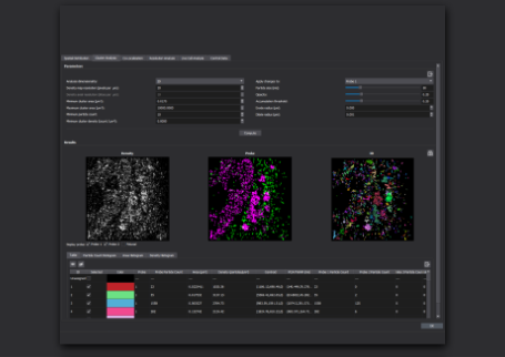
Image Gallery: SMLM Applications in the Neurosciences
Retinal Tissue Slice to Study Neural Development
MAGENTA: CALBINDIN; CYAN: PSD95

Retinal Tissue Slice to Study Neural Development
CALBINDIN

Retinal Tissue Slice to Study Neural Development
PSD95

秀丽隐杆线虫作为突触传播的模型系统

秀丽隐杆线虫作为突触传播的模型系统